Chest X-ray
Chest X-ray
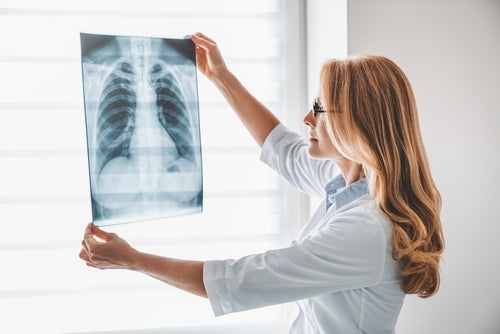
What it is and how it works: A chest x-ray is a quick and painless investigation used to take black and white images of the heart, lungs, and bones. It works by passing x-rays, a type of radiation, through the body to a detector (or photographic plate) at the back. While dense structures such as bones appear white, softer structures such as the heart and lungs appear dark. Sometimes, images will be taken from different angles to better visualise the organs of the body. Usually, the whole process will only take a few minutes.
What it detects: A chest x-ray can be requested if a doctor thinks that the cause of your symptoms might be due to a lung or heart issue. It can help to detect problems with the lungs, such as pneumonia or lung cancer, or problems with the heart, such as heart failure. It can also be done as part of routine work up for an operation.
